
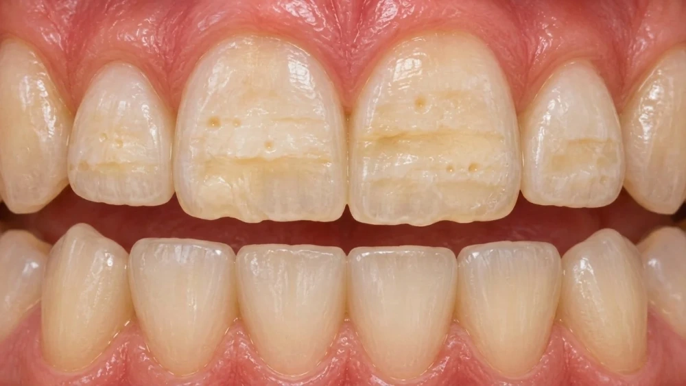
Enamel hypoplasia is a condition in which the tooth’s protective outer layer did not fully form. Perhaps a tooth has always had a groove across it, or tiny pits keep collecting stain despite careful brushing.
Hypoplastic dental enamel can affect children and adults, sometimes on a single tooth and sometimes across several. It is not simply a sign of poor brushing. The defect develops while the tooth is forming, before it appears in the mouth.
The missing enamel cannot grow back, but there are ways to protect affected teeth, manage sensitivity and improve their appearance.
What is hypoplastic dental enamel?
Enamel hypoplasia is a developmental defect that leaves a tooth with less enamel than it should have. The enamel may be thin in certain areas or missing altogether, creating pits, grooves or an uneven outline.
Dentists describe this as a quantitative defect: the amount of enamel is reduced. The enamel that remains is not necessarily soft. Both baby teeth and permanent teeth can be affected. The location and extent depend partly on which teeth were developing when enamel formation was disrupted.
Is it the same as enamel erosion?
No. Hypoplasia starts during tooth development. Erosion is the chemical loss of tooth tissue after teeth have erupted, for example through repeated exposure to acids.
An adult may only recently notice a developmental defect that has been present for years. However, new surface damage can also come from decay, erosion or wear, so it should not automatically be labelled hypoplasia.
What does enamel hypoplasia look like?
The most useful clues are changes in the tooth’s surface and shape. Signs may include:
- Small pits or depressions.
- Horizontal grooves or bands of reduced enamel.
- Areas where the enamel is noticeably thin or absent.
- Yellow or brown areas where underlying dentine shows through or staining collects.
- Sensitivity, particularly where the tooth has less protection.
Not everyone has pain, and not every discoloured patch is hypoplasia. White spots can occur with hypomineralisation, fluorosis or early decay as well. Colour alone cannot establish the diagnosis.
If a newly erupted tooth already looks unusual, arrange a dental examination rather than trying to scrub or whiten the mark away.
What causes enamel hypoplasia?
Enamel is produced by specialised cells called ameloblasts. A disturbance during the stage when they lay down the enamel matrix can leave too little enamel behind. Possible explanations include inherited conditions, local injury and disturbances in general health during development.
Inherited enamel conditions
Amelogenesis imperfecta is a group of genetic disorders affecting enamel formation. Its hypoplastic forms produce thin enamel, while other forms mainly affect hardness or maturation. It may involve both baby and permanent teeth.
Amelogenesis imperfecta is one possible cause of hypoplasia, not another name for every case. A family history of similar teeth can be helpful, although its absence does not exclude a genetic explanation. Widespread defects may need specialist assessment, particularly where other dental or medical findings are present.
Injury or infection near a developing tooth
Trauma to a baby tooth can affect the permanent tooth developing beneath it. Infection around a baby tooth is another possible local disturbance. The permanent tooth may later emerge with a defect even though the original incident happened years earlier.
An isolated defect associated with a local disturbance is sometimes called a Turner’s tooth. The appearance depends on the timing and severity of the injury to development.
Health and nutritional disturbances
Significant illness, nutritional deficiencies and certain medical conditions during tooth development have been linked with enamel defects. The effect depends on when the disturbance occurred and which teeth were forming.
Research also links premature birth and low birth weight with a higher risk of enamel hypoplasia in baby teeth. This is an association across groups of children, not a prediction for an individual child. Many children born prematurely will not have the same dental findings, and an enamel defect does not establish why a pregnancy or birth was complicated.
These associations do not mean a tooth mark proves a particular vitamin deficiency, illness or problem during pregnancy. Often, the exact cause cannot be established. Taking supplements later will not replace enamel that failed to form.
Enamel hypoplasia vs hypomineralisation
These terms are often confused because both conditions can cause discolouration and damaged-looking teeth. The difference lies in how the enamel developed.
Feature | Enamel hypoplasia | Enamel hypomineralisation |
Main problem | Too little enamel formed | Enamel did not mineralise normally |
Initial thickness | Reduced in affected areas | Often normal when the tooth erupts |
Typical appearance | Pits, grooves or missing areas | Defined or diffuse patches of altered colour, depending on the condition |
What happens later | Thin or uneven areas may be vulnerable to damage | Weaker enamel may break down after eruption |
Once enamel has broken away, the original problem can be harder to identify. A dentist considers the pattern and history as well as the current appearance.
Molar-incisor hypomineralisation (MIH) is a particular developmental enamel condition affecting one or more first permanent molars, sometimes with permanent incisors. It is not another name for enamel hypoplasia.
How is enamel hypoplasia diagnosed?
The dentist examines the teeth for reduced enamel thickness, surface defects, decay and signs of breakdown. They may ask whether the changes were present when the teeth erupted, whether baby teeth were affected and whether relatives have similar problems.
Previous injury, childhood illness and relevant medical history can help explain the pattern. Photographs or older dental records may also be useful. Where indicated, X-rays can help assess enamel thickness, its contrast with dentine and any associated disease. Genetic testing may be considered if an inherited enamel disorder is suspected; it is not needed for every isolated pit or groove.
The distribution offers clues. One affected tooth may suggest a local event, whereas similar defects on teeth that developed at the same time may point towards a more general disturbance. These patterns help guide the investigation; they cannot identify a cause with certainty on their own.
The diagnosis guides treatment: a stain, an early cavity and an area of missing developmental enamel do not necessarily need the same approach.
Enamel hypoplasia treatment: what can help?
Treatment protects the tooth that remains and restores damaged areas where necessary. The choice depends on symptoms, the amount of missing tissue, the bite, age and appearance concerns.
Treatment | Main purpose | Important limitation |
Fluoride and preventive care | Reduce decay risk and help protect existing enamel | Cannot recreate missing enamel thickness |
Sealants | Protect suitable pits and grooves | Need monitoring and may not suit extensive defects |
Composite bonding or fillings | Rebuild local defects and improve shape | May need maintenance or replacement |
Protect teeth with substantial structural damage | More extensive treatment; selected according to need and age | |
Improve selected adult front teeth with visible defects | Require suitable bonding conditions and careful planning | |
Microabrasion or resin infiltration | Improve selected superficial discolouration or enamel opacities | Do not replace substantial missing tooth structure |
Does every affected tooth need treatment?
A shallow, symptom-free defect may need monitoring and extra prevention rather than drilling. A deeper pit that collects plaque, an area of exposed dentine or a tooth that is breaking down presents a different problem.
Treatment is often staged: first make eating and cleaning comfortable, then control any decay and restore vulnerable areas. Cosmetic changes can follow once the teeth are stable. Ask your dentist what each proposed procedure is intended to achieve and whether a less extensive option could do the job.
Prevention and sensitivity management
Fluoride toothpaste, professionally applied varnish and an individual preventive plan can help protect teeth. Sensitivity may improve with appropriate products or by covering exposed areas. Persistent pain needs examination rather than repeated use of a desensitising toothpaste alone.
Remineralisation supports existing tooth tissue; it does not reverse the developmental absence of enamel. Products advertised as “enamel repair” should be understood in that context.
Bonding, veneers and crowns
Composite resin can fill pits, rebuild shape and cover some discoloured areas. Where damage is more extensive, a crown may provide protection. For back teeth, zirconia crowns offer high strength, while emax crowns suit front teeth where natural translucency matters. Veneers can be considered for selected adult front teeth, but suitability depends on how much reliable tooth tissue is available for bonding.
The aim is to preserve sound tissue while providing a restoration that can function well. A mild defect does not automatically justify a veneer or crown.
Restorations also need looking after. A bonded area may require polishing or repair, and a crown or veneer may eventually need replacement. Discuss that longer-term commitment alongside the immediate result, especially if several front teeth are being considered.
What about whitening and white-spot treatments?
Whitening changes colour; it does not fill a groove or restore thickness. It can cause sensitivity, so professional assessment is particularly useful when enamel is already thin.
Microabrasion removes a small amount of surface enamel and requires careful selection. Resin infiltration may help the appearance of certain opacities, but cannot rebuild a missing section. Results reported for selected cases should not be treated as a universal solution for hypoplastic teeth.
Looking after hypoplastic teeth at home
For children under three, NHS guidance recommends a smear of toothpaste containing at least 1,000ppm fluoride unless the dentist advises otherwise. Children aged three to six use a pea-sized amount of 1,000–1,500ppm toothpaste. Parents should help or supervise brushing and discourage swallowing toothpaste.
Do not file rough areas yourself or use acidic DIY whitening mixtures. If brushing hurts, ask for help managing the sensitivity so cleaning remains possible.
- Brush twice daily with fluoride toothpaste. Use a soft brush and avoid aggressive scrubbing.
- Clean between teeth daily with an appropriate method recommended by your dental team.
- Reduce frequent sugary snacks and drinks. Repeated exposure increases the opportunity for decay.
- Limit frequent acidic drinks and discuss reflux or other sources of acid exposure if relevant.
- Attend reviews at the interval advised. Your risk and restorations determine how often checks are needed.
When should you see a dentist?
Book an examination for visible pits, grooves or defects on newly erupted teeth, persistent sensitivity, chipping or a changing discoloured area. Seek prompt care for severe pain, swelling or signs of infection, which may require root canal treatment.
For a child, an early examination gives the dentist a chance to protect affected teeth before problems accumulate. The NHS recommends a first visit when the first baby teeth appear or before the first birthday.
FAQs
Can enamel hypoplasia be cured?
Missing developmental enamel cannot regrow. However, prevention and restorations can protect teeth, improve comfort and restore their appearance. The amount of treatment varies considerably between people.
Is enamel hypoplasia caused by poor brushing?
No. It develops before the tooth erupts. Brushing and diet influence what happens afterwards, including the risk of decay, but they did not cause the original developmental defect.
Does hypoplasia in baby teeth mean adult teeth will be affected?
Not necessarily. Baby and permanent teeth form at different times, and a local disturbance may affect only one tooth. Some inherited conditions affect both sets. A dentist can assess the pattern and monitor permanent teeth as they emerge.
Does enamel hypoplasia get worse with age?
The original formation defect does not spread, but affected teeth can develop decay, wear or fractures over time. Changes in symptoms or appearance warrant a review.
Can enamel hypoplasia be prevented?
Not every cause can be prevented, particularly inherited conditions. Appropriate medical care, nutrition and prompt attention to injured or infected baby teeth support health, but cannot guarantee normal enamel formation. Once an affected tooth erupts, prevention focuses on avoiding additional decay and wear rather than changing how that tooth developed.
Can adults develop enamel hypoplasia suddenly?
An adult may notice it for the first time, but the defect formed during development. Sudden changes in previously normal-looking teeth need assessment for other causes, including decay, erosion and wear.
Resources
- Cleveland Clinic. “Enamel Hypoplasia.”
https://my.clevelandclinic.org/health/diseases/enamel-hypoplasia - Patel A, Aghababaie S, Parekh S. “Hypomineralisation or hypoplasia?” British Dental Journal. 2019;227:683–686.
https://pubmed.ncbi.nlm.nih.gov/31654000/ - MedlinePlus Genetics, National Library of Medicine. “Amelogenesis imperfecta.”
https://medlineplus.gov/genetics/condition/amelogenesis-imperfecta/ - Reynolds L, Dave M, Tattar R, Barry S. “Inherited dental anomalies – part 1: enamel defects.” Faculty Dental Journal. 2024;15(3).
https://publishing.rcseng.ac.uk/doi/10.1308/rcsfdj.2024.31 - American Academy of Pediatric Dentistry. “Pediatric Restorative Dentistry.”
https://www.aapd.org/research/oral-health-policies–recommendations/pediatric-restorative-dentistry/ - National Health Service. “Children’s teeth.”
https://www.nhs.uk/live-well/healthy-teeth-and-gums/taking-care-of-childrens-teeth/ - National Institute of Dental and Craniofacial Research. “The Tooth Decay Process: How to Reverse It and Avoid a Cavity.”
https://www.nidcr.nih.gov/health-info/tooth-decay/more-info/tooth-decay-process - Guy’s and St Thomas’ NHS Foundation Trust. “Molar incisor hypomineralisation in children.”
https://www.guysandstthomas.nhs.uk/health-information/molar-incisor-hypomineralisation-children - Caeiro-Villasenín L et al. “Developmental Dental Defects in Permanent Teeth Resulting from Trauma in Primary Dentition: A Systematic Review.” International Journal of Environmental Research and Public Health. 2022;19(2):754.
https://pubmed.ncbi.nlm.nih.gov/35055575/ - Olczak-Kowalczyk D et al. “Developmental enamel defects and their relationship with caries in adolescents aged 18 years.” Scientific Reports. 2023;13:4932.
https://pubmed.ncbi.nlm.nih.gov/36973358/ - Oliveira A et al. “Dental Bleaching, Microabrasion, and Resin Infiltration: Case Report of Minimally Invasive Treatment of Enamel Hypoplasia.” International Journal of Prosthodontics. 2020;33(1):105–110.
https://pubmed.ncbi.nlm.nih.gov/31860920/ - Xu S et al. “Relationship between preterm, low birth weight, and development defects of enamel in the primary dentition: A meta-analysis.” Frontiers in Pediatrics. 2022;10:975340.
https://pubmed.ncbi.nlm.nih.gov/36440332/ - Kanchan T, Machado M, Rao A, Krishan K, Garg AK. “Enamel hypoplasia and its role in identification of individuals: A review of literature.” Indian Journal of Dentistry. 2015;6(2):99–102.
https://pmc.ncbi.nlm.nih.gov/articles/PMC4455163/